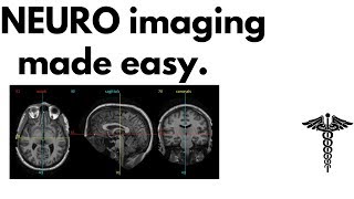
Cases in Radiology: Episode 1 (neuroradiology, CT, MRI)
This episode takes you through an intermediate difficulty neuroradiology case complete with CT, MRI and histology. View the case in the Radopaedia quiz mode ...
Radiology Channel
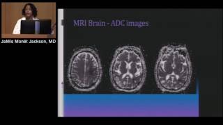
Neuroradiology: Basic Interpretation of CT & MRI (JaMís Jackson, MD)
DeBakey Institute for Cardiovascular Education & Training (DICET) RADIOLOGY FOR THE NON-RADIOLOGIST Neuroradiology: Basic Interpretation of CT ...
Houston Methodist DeBakey CV Education
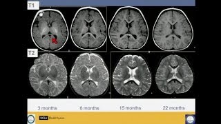
How to Read and Interpret Pediatric Brain Tumor Imaging Reports
Dr. Michael Fisher offers parents tips to interpret child's imaging results and what the reports mean.
Penn Medicine
Neuroradiology physics review - 1 - Computed Tomography
It's important for the neuroradiologist to have a basic grasp of physics, particularly in the ways that it may affect image quality. In this video, Dr. Michael Hoch ...
LearnNeuroradiology
Imaging of Brain tumors (New WHO Classification) - Prof Dr. Mamdouh Mahfouz
Imaging of Brain tumors based on (New WHO Classification) discussed by Prof Dr. Mamdouh Mahfouz (In Arabic)
Mamdouh Mahfouz
Imaging of Brain tumors - DRE 3 - Dr Mamdouh Mahfouz
Imaging of Brain tumors - DRE 3 - Dr Mamdouh Mahfouz.
Mamdouh Mahfouz
Space Occupying Lesions Part 1
Credit: Mueez Waqar www.nansig.org.
NANSIG
CT v MRI brain
This project was created with Explain Everything ™ Interactive Whiteboard for iPad.
Steve Jacques
Head CT Interpretation Made Easy
This is a lecture given to emergency medicine providers discussing how to read a head CT. It was given on 9/26/15 by Dr. Hartmut Gross at the Rural Emergency ...
Daniel McCollum
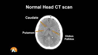
Normal Head CT Scan Anatomy Made Simple- Neuroradiology
This video is a part of basic radiologic head CT SCAN anatomy series. The video shows the basic CT anatomy of the brain. For each slice we have highlighted ...
Medcrine Medical
Introduction to the CT Brain
Dr. Charif Sidani demonstrates his approach to interpreting the Brain CT.
Radiology Residency UM/JMH
WEBINAR: Reading Your Radiology Report
Radiologists use medical imaging to help physicians diagnose, treat, and monitor brain tumors. Join this webinar to learn how to understand and interpret your ...
American Brain Tumor Association
CT Brain -Checklist
Approach to CT Brain interpretation and final checklist.
Shades of Radiology
Neuroimaging of Pediatric Disease
Visit: http://www.uctv.tv/) Professor of Radiology from the David Geffen School of Medicine UCLA, Noriko Salamon, MD, PhD, reviews basic imagery techniques ...
University of California Television (UCTV)
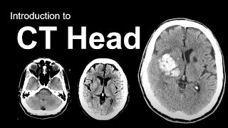
Introduction to CT Head: Approach and Principles
Video includes relevant anatomy (4:50), basic principles, approach to CT head (38:00), and multiple example cases (41:54). The goal of this video is to teach ...
Navigating Radiology
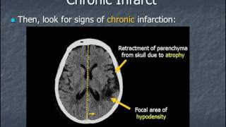
Identifying early computed tomography (CT) findings of infarctions
After viewing this video, from our Brain CT Essentials course, you will be able to recognize the early computed tomography (CT) findings of an infarct. ⏯Start the ...
Medmastery
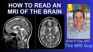
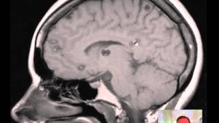
How to Read an MRI of the Brain | First Look MRI
Dr. Brian Gay provides an easy to understand explanation of an MRI brain scan and how to read it. With the premium service, First Look MRI provides a video ...
First Look MRI
Introduction to Brain Surface Anatomy
Speaker: Dr. Balaji Rao, MD. Assistant Professor of Radiology and Biomedical Imaging, Yale University School of Medicine.
Yale Radiology and Biomedical Imaging
Clinical trends and new tracers for Neuro-PET/CT
Clinical trends and new tracers for Neuro-PET/CT. Presented by Torsten Danfors, MD PhD Section for Nuclear Medicine and PET, Department of Surgical ...
GE Healthcare
Imaging of Brain tumors Dr Mamdouh Mahfouz
Brain tumors Dr Mamdouh Mahfouz.
Mamdouh Mahfouz
Intro to Head CT Part II: Evaluation of Ischemic Stroke
A Division of Hospital Medicine Grand Rounds presented by Puneet Pawha, MD, Division of Neuroradiology.
Icahn School of Medicine
Brain Anatomy MRI- Neuroradiology
This video shows the appearance of the anatomical structures of the brain on a Magnetic Resonance Imaging. It aims to complement your understanding of ...
Medcrine Medical
Image Guided Surgery for Brain Tumors
Dr. Chris McPherson explains how image-guided surgery (IGS) technology is used in the operating room. Similar to a GPS for your car, an IGS system helps the ...
Mayfield Brain & Spine
Pre-operative Imaging - Craniopharyngioma | UCLA Pituitary Tumor Program
UCLA Health
AN APPROACH TO CT HEAD NARRATED 2
James Kinkaid
Neuroanatomy on MRI | Part 1| Cerebrum, Basal Ganglia, Thalamus, Internal Capsule & Lesions
Hello again doctors! This is part 1 of high yield neuroanatomy by MRI and 3D model. I go over the axial slices that are above the brainstem and go over the ...
Slay Step 1
Bilateral Parotid Diseases - Radiology Review Course (MRI, CT, ultrasound)
A short extract from the Head & Neck section of Radiopaedia's Radiology Review Course. You can purchase access to the full 6 hour video course on our ...
Radiology Channel
Intracranial infections - 3 - Focal Infections
Focal infections are those infections of the brain which are walled off either within the brain parenchyma or in the extra-axial space, such as subdural or epidural ...
LearnNeuroradiology
Radiological anatomy of Brain - DRE 1 - Prof. Dr. Mamdouh Mahfouz
Radiological anatomy of Brain - DRE 1 - Prof. Dr. Mamdouh Mahfouz.
Mamdouh Mahfouz
How to Read Brain MRI by Dr Hamza Alsayouf
Brain MRI cuts and anatomy.
American Child Neurology Center of UAE
Brain tumor ct
Mass in brain ct scan.
CT pathology
Cysticercosis (multiple) of Brain MRI DISCUSSION
This is a discussion of an MRI of the brain in a 30yoM with Numerous cysticercosis lesions of the brain.
rdavidm1
Imaging features of meningiomas - Part 1
Speaker: Dr. E. Leon Kier, MD. Professor of Radiology and Biomedical Imaging, Yale University School of Medicine.
Yale Radiology and Biomedical Imaging
Imaging of Brain tumors - Dr Mamdouh Mahfouz (In Arabic)
Imaging of Brain tumors - Dr Mamdouh Mahfouz (In Arabic) Neurology conference ( Feb 2014 ) at Menia city.
Mamdouh Mahfouz
Radiology: How to Read a CT Abdomen & Pelvis (My search pattern)
Ever wonder how a RADIOLOGIST reads a CT Abdomen + Pelvis? This is a quick overview of the search pattern I use for every CT Abdomen & Pelvis I read.
Dr. Cellini
A non-contrast CT for a patient with focal neurological signs and symptoms
Mini Been Non-Contrast CT Scan is the initial diagnostic test in a patient with focal neurological signs and symptoms with an unknown or unclear history.
Drbeen Medical Lectures
A neurologist's perspective on the benefits of digital PET imaging
A neurologist's perspective on the benefits of digital PET imaging. Presented by Dr. Stephane Epelbaum, Pitié Salpêtrière Hospital, Paris, France.
GE Healthcare
Extra axial lesion - Easy Signs to identify
Simplified approach to extraxial lesions with classic examples.
Shades of Radiology
Imaging of Brain tumors - Prof Dr. Mamdouh Mahfouz (mans2017)
Imaging of Brain tumors discussed by Prof Dr. Mamdouh Mahfouz (In Arabic)
Mamdouh Mahfouz
Christopher Hess, MD, PhD, Neuroimaging Part 2: Fundamentals of Image Interpretation
The easiest way to seperate an MRI from a CT scan is to look at the outside of the head. CT has little tissue contrast, but the bone is bright. With an MRI, tissue ...
UCSF School of Medicine
Demyelination Case Review - ADC Negative Multiple Sclerosis
MRI Mastery Series: Demyelinating Disease by Dr. David Yousem, MD - https://mrionline.com/p/mri-mastery-series-demyelinating-disease/ Dr. Yousem is back ...
MRI Online
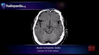
Stroke: Acute infarction - radiology video tutorial (CT, MRI, angiography)
"Stroke Series" video 3 of 7: Acute ischaemic stroke. Presented by Neuroradiologist Dr Frank Gaillard. Find out more: ...
Radiology Channel