how to read mri LS spine
CT Scan & MRI
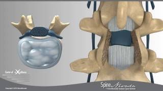
Lumbar Disc Nomenclature on MRI
How to name lumbar disc herniations properly and according to a consensus paper by the combined task forces of the North American Spine Society, the ...
Dr Christoph Agten
How To Read Lumbar Spine MRI: Axial Anatomy
Review of difficult to understand axial anatomy on Lumbar Spine MRI. By Sarel Gaur MD.
Sarel Gaur MD
VERTEBRAL COLUMN ANATOMY (1/2)
Your spine, also called your backbone or vertebral column, is composed of 33 bones, called vertebrae, which provide your body with support and protect your ...
Neural Academy
How to Read a MRI of the Normal Thoracic Spine (Mid Back) | Vail Spine Specialist
For Patients: http://neckandback.com For Professionals: http://studyspine.com Accredited Training: http://studyspine.com/register/ Forum Discussion: ...
Donald Corenman, MD, DC
How to Read a Cervical Spine MRI Part 1: An Anatomy Overview and Focus on the Degenerative Spine
Learning Objectives: 1. Review relevant CT and MRI cervical spine anatomy. 2. Discuss common chronic cervical pain generators and how they may be caused ...
ASM
How to read and MRI of the cervical spine
The video describes the cervical spine anatomy and the approach to reading a cervical spine MRI by Dr. Gay, a radiologist.
First Look MRI
How to Read a Spine MRI
In this video, Dr. Webb goes over (in basic terms) how to read a lumbar MRI. MEDICAL DISCLAIMER The information provided is not intended to be a substitute ...
Antonio J. Webb, M.D.
School of Medical Imaging MRI Lab - T spine
Randy Paquette
Spinal Cord - Clinical Anatomy and Physiology (dermatomes, blood supply, shingles, lumbar puncture)
Buy Images here: armandoh.org/shop Where do I get my information from: http://armandoh.org/resource Facebook: ...
Armando Hasudungan
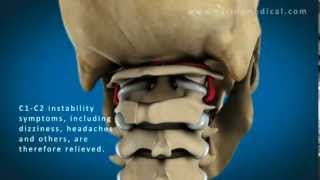
C1 and C2 Atlantoaxial Instability
This video is an animated demonstration of C1-C2 instability and is not meant to be an exact anatomical model. Atlantoaxial instability or upper cervical ...
Caring Medical Regenerative Medicine Clinics
Thoracic Spine Anatomy and Palpation with Michael Lucido
View and excerpt form the C-613 Thoracic Spine course with NAIOMT Faculty Member Michael Lucido as he goes over thoracic spine anatomy and palpation.
NAIOMT1
MRI Anatomy of the HIP
Dr. Jean Jose reviews the detailed anatomy of the Hip/pelvis. Muscles, bones and joint structures are reviewed in detail with tidbits of key points required for the ...
Radiology Residency UM/JMH
Dr. Gillard lectures on How to Read Your Lumbar MRI
In this video, Dr. Douglas Gillard explains the basics of how to read your lumbar MRI. You will learn the difference between a sagittal versus axial image, and ...
Douglas Gillard, BS, DC, Spine Researcher
Vertebral Column – Anatomy | Lecturio
This video “Vertebral Column” is part of the Lecturio course “Abdominal Wall - Anatomy” ▻ WATCH the complete course on http://lectur.io/vertebralcolumn ...
Lecturio Medical
How to Read a MRI of the Normal Cervical Spine (Neck) | Colorado Spine Expert
For Patients: http://neckandback.com For Professionals: http://studyspine.com Accredited Training: http://studyspine.com/register/ Forum Discussion: ...
Donald Corenman, MD, DC
How to read an MRI
Dr. John Shim of Shim Spine gives an overview of how to read MRI imagery. www.shimspine.com For more information on this subject, please see our blog at ...
Shim Spine
Normal Cervical Spine MRI Explained | Dr. Jeffrey P.Johnson | HD
In this video Dr. Jeffrey P. Johnson demonstrates the normal appearance and anatomy of the cervical spine as seen on an MRI scan. Click on our channel for ...
PortlandNeurosurgery
Medical imaging at the level of C1: CT and MRI (preview) - Radiological Anatomy | Kenhub
At the level of the 1st cervical vertebra, there are many important structures, like muscles and bones that can be seen using CT and MRI. Watch the full video ...
Kenhub - Learn Human Anatomy
Anatomy of the Spine - NUCLEUS Medical Media
UPDATE, 11/2011: A new version of this animation is now available! Watch it here: https://youtu.be/KiriyiVZae8 This animation shows the anatomy of a typical ...
Nucleus Medical Media
how to read neck mri axial
CT Scan & MRI
the spine w mri
SCC Pathology
Weight bearing MRI provides optimal diagnosis of lumbar disc herniation - Reno, Sparks, Carson
The tiltable Esaote G-Force Brio at SpineNevada provides more detail, better accuracy and greater confidence. For conditions that involve nerve root ...
SpineNevada
Weight-Bearing MRI of the lumbar spine
Clinical implications of Weight-Bearing MRI of the lumbar spine and the potential to improve patient treatment and outcome. Courtesy of: Dr. Mikael Boesen ...
esaotegroup
Up-right MRI of the lumbo-sacral spine: 10 years after
Courtesy of: Prof. Massimo Gallucci - Professor and Chairman of Neuroradiology, University of L'Aquila - Head of Neuroradiology Unit - S. Salvatore Hospital, ...
esaotegroup
SI Joint Anatomy, Biomechanics & Prevalence
To speak with a nurse or find a surgeon call 888-689-5277 or visit http://si-bone.com/patients/next-steps/find-a-doctor/ Learn more about the sacroiliac joint at ...
SI-BONE
Spine Lecture: How to Read a Cervical MRI
Upper Chesapeake Medical Center Spine Conference 5/1/15.
Spiro Antoniades, M.D.
Demonstration of weight-bearing MRI revealing lumbar stenosis: SpineNevada Sparks, Reno, Carson City
Lumbar spinal stenosis can often cause increased pain, numbness and/or weakness while standing or sitting. The symptoms may be relieved when lying down.
SpineNevada
Multi-position, open MRI reveals spine disorder - Carolina Neurosurgery & Spine Associates
Patient Tracy Bari found relief from intense back pain by undergoing minimally invasive lumbar fusion (TLIF). The on-site multi-position, open MRI at Carolina ...
Carolina Neurosurgery & Spine
Dr. Casden Performs Anterior Cervical Spine Surgery
In this video segment, Andrew Casden, MD, describes the history and MRI of a patient with a cervical disc herniation and performs the surgery with a descriptive ...
Mount Sinai Health System
Anatomy of a Coronal MRI of the Thorax
An annotated overview of the anatomy visible in a coronal Gradient echo T1 (fat supressed) magnetic resonance image of the thorax (and part of the abdomen).
Eva Sweeney
Diagnosing Neck Pain with MRI's
Every year millions of people experience neck and upper extremity symptoms and do not know the seriousness of these conditions. Gerard A. Compito, MD, ...
PrincetonHealth
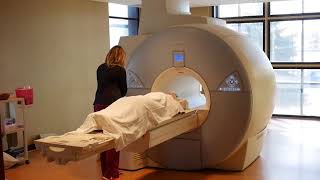
What to Expect from an MRI
Experiencing pre-exam jitters? This video walks you through every step of an MRI exam: https://goo.gl/5zW2qN #WeSpecializeInAnswers.
Center for Diagnostic Imaging
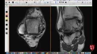
Systematic Interpretation of Ankle MRI: How I do it
Introductory, yet detailed review of ankle anatomy on MRI to serve as a foundation for clinical case review. Labelled anatomy of the ankle is available at ...
Chris Beaulieu
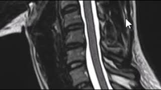
Spinal Cord Compression
Overview of spinal cord compression including pathophysiogy, symptoms, diagnosis, and management. To learn more about spinal cord compression, visit ...
Learn Oncology
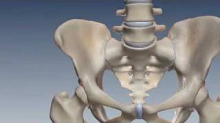
Where is the Sacroiliac Joint? Anatomy of the Sacroiliac Joint
The last segment of the spine is the sacrum. The sacrum attaches to the large pelvic bones known as the ilium. The joints that connect the sacrum to the ilium are ...
SI-BONE
Ultrasound of the Neonatal Head and Spine
Webinar recording for Ultrasound of the Neonatal Head and Spine presented by Dr Sheryle Rogerson. The topics covered include clinical indications through to ...
GE Healthcare
Q-Spine, the next step in MRI spine analysis
Q-Spine is a support tool for Visualization and Quantification of relative biomechanical changes of the spine comparing Weight-Bearing and Recumbent MRI ...
esaotegroup
How to Read Your MRI with Onis 2.5 - part 2 of 2 (Advanced Lumbar Spine MRI Anatomy)
In this video Dr. Gillard demonstrates how to use Onis 2.5 (a free MRI viewing software) to view a real MRI disk. He goes over basic MRI anatomy on a split ...
Douglas Gillard, BS, DC, Spine Researcher
Low Back Pain and the Sacroiliac Joint - Dr. Alexander
An important but often overlooked cause of low back pain may be the sacroiliac (SI) joint. Unresolved low back pain, hip, groin or leg pain may be coming from ...
SI-BONE
Lumbar Laminectomy and Fusion Presented by Spine Nevada, Reno Spine Surgeons and Spine Center
Using a minimally invasive laminectomy, the location of the incision is often established by an intraoperative X-ray, using fluoroscopy. A skin incision about 1 ...
SpineNevada
Gray and white matter | Organ Systems | MCAT | Khan Academy
Created by Matthew Barry Jensen. Watch the next lesson: ...
khanacademymedicine