EPMA Quantitative Analysis Part 1
Sven Holbik
Advanced quantitative fluorescence microscopy to probe the molecular dynamics of viral entry
Presented By: Luis Alvarez, Ph.D. Speaker Biography: Dr. Luis Alvarez studied physical chemistry at the Université Paris-Sud XI in Orsay where he worked on ...
LabRoots
Multi-colour biological electron microscopy using EDS
Presented By: Louise Hughes Speaker Biography: Louise is the Product Manager for Life Sciences at Oxford Instruments NanoAnalysis. Louise specialises in ...
LabRoots
AFM Enhancing Traditional Electron Microscopy Applications | Bruker Webinar
This webinar takes a look at recent AFM technical advances that provide a highly productive nano-imaging workflow, akin to current electron-microscopy based ...
BrukerNanoSurfaces
Quantitative Imaging of Living Biological Samples by PeakForce QNM Atomic Force Microscopy | Bruker
The promising PeakForce QNM® imaging mode and Peak Force Tapping™ technique applied by the BioScope™ Catalyst™ AFM microscope look to provide ...
BrukerNanoSurfaces
Electron Holography I - Prof. Etienne Snoeck
Aula ministrada pelo Prof. Etienne Snoeck, durante a sexta edição do Transmission Electron Microscopy (TEM) Summer School, realizado pelo Laboratório ...
LNNano
Lecture 10: Quantitative metallography
This lecture discusses the different methods used to determine the grain size of a microstructure.
Material Science and Engineering - IITR
MiniTEM - low voltage electron transmission microscope
MiniTEM is designed for sub-visible particle characterization and is typically used for automated purity analysis in biopharmaceutical development and ...
VironovaAB
In situ transmission electron microscopy: formation of α
Tracking the αʺ martensite decomposition during continuous heating of a Ti-6Al-6V-2Sn alloy Pere Barriobero-Vila, Verona Biancardi Oliveira, Sabine Schwarz, ...
TITANIUM
EPMA Quantitative Analysis Part 2
Sven Holbik
Gian@JNU: Advanced Electron Microscopy for Materials Science-P11
Prof. Dr. Benjamin Butz, University of Siegen, Germany 13-22 March 2018.
GIAN JNU
Electron Microscopy (Lecture No 8)
PLANET OF BOTANY
Introduction to Electron Probe X-Ray Microanalysis (EPMA) by Dr Jeff Chen
This webinar is presented by Dr Jeff Chen from the Centre for Advanced Microscopy at Australian National University, a Microscopy Australia facility.
Microscopy Australia
Applications of Deep Learning in Astronomy and Electron Microscopy - Ajit Kembhavi - 6/25/2019
AstroInformatics 2019 Conference: AstroInformatics Methods and Applications http://astroinformatics2019.org/
caltech
let's Talk Science: What, Why, and How? Session on "Electron Microscopy and Spectroscopy"
Let's listen to Dr. A.K. Srivastava on "Electron Microscopy and Spectroscopy" over CSIR-JIGYASA: Student-Scientist Connect Programme as Scientific Social ...
CSIR-AMPRI, Bhopal
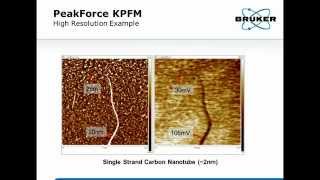
High Resolution Quantitative Kelvin Probe Force Microscopy - Principles and Applications | Bruker
In our new probe and instrument designs, we have found ways to reduce probe to probe measurement scatter to below a standard deviation of 50 mV.
BrukerNanoSurfaces
Scanning electron microscopy/EDS
prof.kashif samoo and rafiq pitafi.
Rafique pitafi lectures and experiences
NACK Course Notes: Electron Microscopy
NACK Network: nano4me.org Educator Resources: http://nano4me.org/educators.
Nack Center
ZEISS: Your Complete Microscopy Solution for Oil & Gas
ZEISS offers multi-scale imaging from centimeter to nanometer in a single, automated environment for visible petrography & quantitative mineralogy. Perform 3D ...
ZEISS Microscopy
EPMA Quantitative Analysis Part 4
Sven Holbik
UPenn MEAM Department Seminar - In Situ Liquid Cell Electron Microscopy with the NanoAquarium
More information on the nanoAquarium here: http://bau.seas.upenn.edu/research/in-situ-electron-microscopy-of-liquids-the-nanoaquarium/ Nicholas M ...
Nicholas Schneider
11 - Introduction to electron diffraction in TEM
Why doing diffraction in TEM and what g-vector is are explained.
Kelvin Xie MSEN TAMU
Scanning Electron Microscope Micro XRF Addition X-Beam.wmv
Micro XRF addition to the Scanning Electron Microscope compliments existing EDS analysis, a whole order of magnitude offering ppm level analysis inside the ...
edskenny
BEHIND THE SCIENCE: In between atoms (National Geographic) (UK)
Take a look between the atoms with the most powerful microscope in the world. And discover, together with the University of Antwerp, how a gold nugget could ...
UAntwerpen
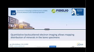
Quantitative backscattered electron imaging, mapping distribution of minerals in the bone specimens
Speaker: Ing. Sofie Kolibova, Ph.D. student at Department of Osteology and Biomechanics, University Medical Center Hamburg-Eppendorf. Title: Quantitative ...
Ondřej Šebesta
Label-Free Quantitative Phase Imaging of Live Cells and Tissue
The development of innovative tools for spatiotemporal imaging of cell and tissue cultures with single cell accuracy significantly advance our understanding of ...
AXT
Microscopy: Fourier Space (Bo Huang)
Learn more: https://www.ibiology.org/talks/fourier-transform/ The Fourier transform is intimately associated with microscopy, since the alternating planes ...
iBiology Techniques
Elementary Stereology for Quantitative Metallography - Course Introduction
NPTEL-NOC IITM
CHArT Seminar Series: High-resolution single-particle cryo-EM
March 12th, 2019 Broad Institute Channel Therapeutics A seminar series with a special interest in ion channels from both biological and therapeutic ...
Broad Institute
BP-ICAM Webinar Series 2019: When Soft Materials Meet Electron Microscopy
In this BP-ICAM webinar, Professor Qian Chen from the University of Illinois at Urbana-Champaign discussed three soft materials systems whose nanoscale ...
The bp International Centre for Advanced Materials
New Frontiers in Cryo Electron Microscopy by Lena Kourkoutis
A seminar presented at the University of Chicago in May 2018.
Center for Bright Beams
Dark Field microscopy in Hindi.
Darkfield_microscope #darkfeild_microscopyin_hinid #THEDRXFAMILY *************************** Tags suggusting this video **************************** Dark ...
The DRx family
Sergei Kalinin: "Deep Learning Dive into the Scanning Transmission Electron Microscopy: Material..."
Machine Learning for Physics and the Physics of Learning 2019 Workshop II: Interpretable Learning in Physical Sciences "Deep Learning Dive into the ...
Institute for Pure & Applied Mathematics (IPAM)
UV-Vis Tutorial | Part 2: Performing a Quantitative Measurement
The second part in a series on how to accurately measure the optical spectra of solutions of nanoparticles using a UV-VIS (UV-Visible) spectroscopy. In this ...
nanoComposix
Electron Microscopy Imaging of Drug Delivery Systems in Liquid State
Speaker: Prof. Dr. Oren Regev, Department of Chemical Engineering, Ben-Gurion University of the Negev (IL) (tbc) CLINAM 7/ 2014, 7th Conference and ...
TAUVOD
Electron Microscopy and X-ray Diffraction -DR Edison H. Ang - EAVERSITY
This video explains the sample preparation and working principles of SEM, TEM and XRD. ✅ Subscribe here for more videos update: ...
EAversity
Phase contrast microscope
Phase-contrast microscopy is an optical-microscopy technique that converts phase shifts in light passing through a transparent specimen to brightness changes ...
Quick Biochemistry Basics
M&M 2020 Presentation: In-Situ Oxidation State Mapping by Electron Energy-Loss Spectroscopy
Liam Spillane, Ph.D. Presentation Number: 652 Session: P06.3 - In-situ TEM at the Extremes - Corrosion Electron energy-loss spectroscopy (EELS) performed in ...
Gatan, Inc.
Yoosuf Picard: Advancing the science of electron microscopy
Professor Picard's research group develops and applies advanced electron microscopy methods for quantitative microstructural characterization and in situ ...
College of Engineering, Carnegie Mellon University
Rostrup Nielsen Symposium 2015 - Stig Helveg: Electron Microscopy in Catalysis
Stig Helveg: Electron Microscopy in Catalysis.
Video History of Catalysis
Phasefocus Virtual Lens
The Phasefocus Virtual Lens® (http://www.phasefocus.com) is a novel method for high fidelity quantitative imaging and microscopy. It is known in the scientific ...
Phasefocus
Interaction between electrons and sample, Imaging capabilities
Materials Characterization by Dr. S. Sankaran Department of Metallurgical & Materials Engineering IIT Madras. For more details on NPTEL visit http://nptel.ac.in.
nptelhrd