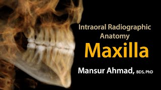
Intraoral Radiographic Anatomy of the Maxilla
Let us learn the radiographic landmarks on the maxillary arch as seen on intraoral radiographs.
Mansur Ahmad
Oral Radiology | Types of Radiographs | NBDE Part II
In this video, we'll cover the different types of radiographs you can take and how to read them: periapical, bitewing, occlusal, panoramic, cephalometric, and ...
Mental Dental
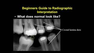
Dental Radiographic Anatomy
Melanie Helmke, RDH, MS.Ed, discusses Dental Radiographic Anatomy.
MATCHEALTH
How to review a manuscript for RadioGraphics
If you are interested in becoming a peer reviewer for RadioGraphics or are already a peer reviewer, this video tutorial featuring Jeffrey Klein, MD, Editor of ...
RadioGraphics
Intraoral Radiographic Anatomy Mandible
Let us learn radiographic anatomy of the mandible as seen on intraoral radiographs.
Mansur Ahmad
Bitewing radiograph
University of Nebraska College of Dentistry Hygiene Class of 2017 demonstrates the paralleling technique of taking intraoral radiographs using phosphor plates.
UNMC College of Dentistry
RADS.110 General Anatomy and Radiographic Positioning Terminology
A beginning video for RADS.110 explaining basic anatomy and radiographic positions and projections.
RadTechEd
Bone Lesions, Radiographic Assessment, Part 2: Classification of Tumors by Geoffrey Riley, M.D.
Review of common bone tumors and their radiographic assessment based on the WHO classification system.
Chris Beaulieu
Extraoral Radiographic Projections | Topics In Description Below
0:00 Introduction 0:18 Lateral Cephalometric 1:49 Submentovertex View 2:02 Canthomeatal Line 3:33 Jug Handle Appearance 4:48 Waters View 6:40 PA ...
MDS Entrance Lectures by Sai Naveen Kumar Pilli
DOTLIB - RSNA Radiographics (Español) - Tutorial
En este video tutorial, conocerá el Journal Radiographics de RSNA, aprenderá a navegar por el contenido disponible, realizará búsquedas básicas y ...
Dotlib TV
Radiographic Positioning
Dental radiographs can be frustrating, time-consuming, and overwhelming. However, if radiography is not performed, 50% of tooth anatomy will be left ...
VeterinaryDentistry
Principles of Radiographic Interpretation
This video is a part of the Oral Radiology Interpretation course at the University of Minnesota School of Dentistry. This video discusses the principles of ...
Mansur Ahmad
Bone Lesions: Radiographic Assessment, Part 1, by Geoffrey Riley, MD
Part 1 of a three part series on assessment of focal bone lesion on radiography. Parts 2 and 3 are in preparation!
Chris Beaulieu
Radiographic Interpretation Section 1
Diane Wilson
Radiographic Image
christyfoster2002
RadioGraphics Publication Information for Authors
This course is directed toward authors of education exhibits at the RSNA Annual Meeting who have been notified that their exhibit has been invited for ...
RadioGraphics
VET Talks - Normal Radiographic Anatomy of the Canine Thorax
VET Talks is a project by the IVSA Standing Committee on Veterinary Education (SCoVE). This VET Talk is by Dr Pete Mantis, DVM, DipECVDI, FHEA, MRCVS, ...
VET Talks
How to Review a PowerPoint submission for RadioGraphics
In this video tutorial, Mindy Horrow, MD, FACR, Section Editor, Women's Imaging for RadioGraphics describes how to review interactive PowerPoint submissions ...
RadioGraphics
VET Talks - Normal Radiographic Anatomy of the Canine Abdomen
VET Talks is a project by the IVSA Standing Committee on Veterinary Education (SCoVE). This VET Talk is by Dr Pete Mantis, DVM, DipECVDI, FHEA, MRCVS, ...
VET Talks
Osseous Radiographic Anatomy of the Upper Extremity
I've put together a quick screencast that demonstrates the osseous radiographic anatomy of the upper extremity. Or, in plain English, I'm going to show you what ...
Douglas Gillard, DC, Professor of Clinical Science
Radiographic Contrast Media (Sanjay Kunapuli, MD) December 4, 2015
Cardiac Catheterization Conference "Radiographic Contrast Media" Sanjay Kunapuli, MD December 4, 2015.
Houston Methodist DeBakey CV Education
Radiographic Interpretation of Pulp and Periapical Infections
This video talks about the distinct radiographic changes in the following lesions and helpful for final year BDS exams: 1. Chronic Pulpitis 2. Acute Apical ...
For the Love of teaching - Dr Shilpi Sharma
Introduction to Spine Radiographs
Speaker: Dr. Balaji Rao, MD. Assistant Professor of Radiology and Biomedical Imaging, Yale University School of Medicine.
Yale Radiology and Biomedical Imaging
RADT 101 Image Formation and Radiographic Quality
christyfoster2002
Plain Film Cardiac and Vascular Anatomy - Frontal Radiograph - Philip Araoz, M.D. - Part 1 of 10
Philip Araoz, M.D., a radiologist at Mayo Clinic, discusses cardiac radiographic anatomy and identifies abnormal cardiac contours on the frontal chest radiograph ...
Mayo Clinic
Maxillary Anterior Periapical Radiograph
University of Nebraska College of Dentistry Hygiene Class of 2017 demonstrates the paralleling technique of taking intraoral radiographs using phosphor plates.
UNMC College of Dentistry
Veterinary Dental Radiographic Interpretation Online Course Preview
Register Here: http://bit.ly/1Hg8Cba for this 5 Hour RACE accredited comprehensive online course detailing veterinary dental radiographic interpretation in dogs ...
VeterinaryDentistry
How to Read an X RAY (Trauma Radiograph) - The Young Orthopod
#fractureclassification #xray #readFractureXRay.
The Young Orthopod
VET Talks- Radiographic Lung Patterns
VET Talks is a project by the IVSA Standing Committee on Veterinary Education (SCoVE). This VET Talk is by Dr Pete Mantis, DVM, DipECVDI, FHEA, MRCVS, ...
VET Talks
Radiographic and Fluoroscopic Equipment
christyfoster2002
Radiographic Positioning of the Skull
My name is Jeremy Enfinger, and I've been teaching for JRCERT-accredited Radiologic Technology programs since 2005. I work full-time in management now ...
Jeremy Enfinger
Plain Film Cardiac and Vascular Anatomy - Frontal Radiograph - Philip Araoz, M.D. - Part 3 of 10
Philip Araoz, M.D., a radiologist at Mayo Clinic, discusses cardiac radiographic anatomy and identifies abnormal cardiac contours on the frontal chest radiograph ...
Mayo Clinic
Reading a chest X-ray
Medical disclaimer: Knowledge Diffusion Inc (DBA Osmosis) does not provide medical advice. Osmosis and the content available on Osmosis's properties ...
Osmosis
How to Read Dental X-Rays
In this introductory video, we talk about how dental x-rays work, how to read them, and how to apply the buccal object rule to localize objects in an x-ray image.
Mental Dental
Imaging of the Hip: More Radiographic Essentials!
An all new introductory lecture on the hip, focused on radiographic analysis of subtle findings. Aimed at trainees in radiology, orthopedic surgery, emergency ...
Chris Beaulieu
VET Talks - How to Read Radiographs
VET Talks is a project by the IVSA Standing Committee on Veterinary Education (SCoVE). This VET Talk is by Dr Pete Mantis, DVM, DipECVDI, FHEA, MRCVS, ...
VET Talks
Medical Student Series: Finding fractures on radiographs
Dr. Danton gives a basic tutorial to give medical students advice on how to approach skeletal radiographs in patients presenting with injury.
Radiology Residency UM/JMH
Nonradiographic Versus Radiographic Axial Spondyloarthritis
Hillary Norton, MD, considers the differences between nonradiographic and radiographic axial spondyloarthritis and discusses the impact of these diseases on ...
AJMCtv
Image Based Discussion | Radiographic Diagnosis
MDS Entrance Lectures by Sai Naveen Kumar Pilli
Digital radiographic image processing
VIDEO INFO: Digital radiographic image processing including histogram analysis, look up table, and various post processing applications. Subscribe! Or we'll ...
Rock The Registry
Radiographic Testing (NDT)
NDT Channel
Maxillary Standard Occlusal Radiograph
One take video instructions on how to take a maxillary standard occlusal radiograph by UNMC College of Dentistry Hygiene Class of 2015 students.
Dr. Shawneen Gonzalez