Tissue Processing for Electron Microscopy
Classic video from Anatomical Sciences, Adelaide University, 2004 Many thanks for all the comments An updated version of this video is now available here ...
medscienceap
Overview of Electron Microscopy analysis in Cellular Pathology at UHL
UHL Genomic Medicine
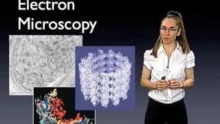
Eva Nogales (UC Berkeley): Introduction to Electron Microscopy
Transmission electron microscopy (TEM) offers the possibility of visualizing biological structures at resolution well beyond that of light microscopy. Whether you ...
iBiology
Chapter 4 (Part 2) Comparison of light and electron microscopy
Overview of types of light microscopy and electron microscopy; Chapter 4 Microbiology by Bauman.
David Singleton
2 The Principle of the Electron Microscope
How to Make a Microscope, Chapter 2 Unlike the optical microscope, the scanning electron microscope uses accelerated electrons in a vacuum to act as light to ...
Thermo Scientific EM & Spectroscopy
Electron microscopy
The Science of Biology
Electron & Light Microscopes | Cells | GCSE Biology (9-1) | kayscience.com
Comparing light and electron microscopes Light microscopes have a lower magnification and resolution than electron microscopes Light microscopes use light, ...
Kay Science
BIOL366L SDSU Electron Microscope Facility Tour
Tour of the San Diego State University Electron Microscope Facility for BIOL 366L.
C1 Biocatalysis
Electron Microscopy for Biological Materials - Kristen Flatt - MRL - 06182020
Electron microscopy is a powerful tool that allows us to overcome the limitations of light to investigate the fine structures that make up the world around us.
MRL Facilities
The Electron Microscope
WinstanleyBiology
Cryo-electron microscopy lab at Goethe-University
The cryo-electron microscopy lab of the Frangakis Group is located at the Goethe Universities Buchmann Institute of Molecular Life Sciences. The main focus of ...
FIAS – Frankfurt Institute for Advanced Studies
An Introduction to Cryo-Electron Microscopy, Dr. Bridget Carragher
CellularImaging #NIGMS #NIH Bridget Carragher, Ph.D., provides an overview of cryo-electron microscopy (cryo-EM). Learn more: ...
National Institute of General Medical Sciences
Renal Pathology : Electron Microscopy and Immunofluorescence Microscopy by Dr. Devesh Mishra.
This Video lesson will be describing about the approach to interpret Electron Microscopy and Immunofluorescence Microscopy of Renal Pathology. --------- To ...
Dr Devesh Mishra
Virtual Tour: Electron Microscopy Imaging
A virtual journey through the Allen Institute for Brain Science's Electron Microscopy (EM) imaging program.
Allen Institute
Scanning Electron Microscopy Facility
Johns Hopkins Applied Physics Laboratory's scanning electron microscopy facility provides a wide range of imaging, analysis and micro-fabrication ...
JHU Applied Physics Laboratory
Electron Microscope (Grade IX Biology)
Smart School System
Electron microscopy lecture | Scanning electron microscope
This electron microscopy lecture explains about the Scanning electron microscopy or SEM principle and advantages. SEM stands for scanning electron ...
Shomu's Biology
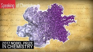
The 2017 Nobel Prize in Chemistry: Cryo-electron microscopy explained
Jacques Dubochet, Joachim Frank, and Richard Henderson have claimed the 2017 Nobel Prize in Chemistry for their development of cryo-electron microscopy.
CEN Online
Ultrastructure of Smooth Muscle Electron Microscopy in Biology and Medicine
Samuel McCombs
Electron microscopy
Tissue preparation and Fixation.
Enas Korina
ZEISS 3D Electron Microscopy for Life Sciences
Cut your samples with an integrated ultramicrotome, slice them with a FIB gun, or image nondestructively with X-ray. Get a maximum of information from your ...
ZEISS Microscopy
Fixative agents used in electron microscopy is
ChandanMLT.
RanKplus
ZEISS Webinar: 3D Electron Microscopy for Life Sciences
Focused ion beam (FIB-SEM) and serial block-face scanning electron microscopy (SBEM) are arising techniques for three dimensional visualization of biological ...
ZEISS Microscopy
Team Talk: Electron Microscopy | Showcase 2017
Researchers from the Allen Institute Electron Microscopy Team present on "Mapping micro-connectivity and cell types using high throughput electron ...
Allen Institute
Electron Microscope (U Bio)
It is important to see what is inside of a cell and its surface but with light microscope it is impossible due to certain limitation. Electron Microscope is very powerful ...
U Bio
Mod-01 Lec-20 Applications of Electron Microscopy
Chemistry of Materials by Prof.S.Sundar Manoharan,Department of Chemistry and Biochemistry,IIT Kanpur.For more details on NPTEL visit http://nptel.ac.in.
nptelhrd
Electron microscope & cell history 9th class
Syeda Anila Waqar
Cryo Electron Microscopy: Revolutionizing the world of structural biology and healthcare
Presented At: Gibco ExpressionWorld 2018 Presented By: Marc Storms - Product Marketing Manager Life Sciences Business Unit, Materials and Structural ...
LabRoots
Osaka University - Research Center for Ultra-High Voltage Electron Microscopy
The development of ultra-high voltage electron microscope and materials and bio-science application of electron microscopy at Osaka University The research ...
WebsEdge Science
Scanning Electron Microscope (SEM) | Principle & Working | GPAT | CSIR NET | GATE | DBT BET | Hindi
In this lecture Scanning Electron Microscope (SEM) discuss in detail with its Principle & Working. At the end of lecture MCQ based on SEM also explained.
Tutor Box
1.2 Resolution of electron microscopes versus light microscopes
Electron microscopes have a much higher resolution than light microscopes as they can resolve to ~0.1nm whereas light microscopes can only resolve to ...
Stephanie Castle
OU Electron Microscopy Facilities
The Microscopy Facility at the Open University campus (Milton Keynes) provides a broad range of light and electron microscopy instrumentation and techniques.
The Open University's Faculty of STEM
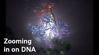
ICR Researchers use Cryo-Electron Microscopy to zoom in on DNA code being read in cells
ICR Researchers led by Dr Alessandro Vannini have captured images of molecular machinery called RNA Polymerase III in the act of transcribing a gene in ...
The Institute of Cancer Research, London
Electron Microscopy Service at Oregon State University
Teresa Sawyer from Oregon State University describes the value of electron microscopy service and why her job is the best in the world.
Thermo Scientific EM & Spectroscopy
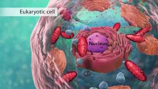
Biology: Cell Structure I Nucleus Medical Media
This animation by Nucleus shows you the function of plant and animal cells for middle school and high school biology, including organelles like the nucleus, ...
Nucleus Medical Media
1.6 What is a Scanning Electron Microscope (SEM) for year 11 biology students
Dr Florence Danila from the Australian National University (anu.edu.au) shows how her work has been directed by the availability of electron microscope ...
ARC Centre of Excellence for Translational Photosynthesis
Cryo-electron Microscopy at the University of Michigan
Three scientists who helped to develop cryo-electron microscopy won the 2017 Nobel Prize in Chemistry. But what is cryo-EM and why is it revolutionizing ...
TheLSIatUM
Using a Scanning Electron Microscope for research
The scanning electron microscope (or SEM) is a microscope that provides a 3D photograph at high magnification levels. It provides up to 300 thousand times ...
NationwideChildrens
Use of a Table Top Scanning Electron Microscope FINAL
Advanced Taxonomic Training Videos
Advantages and disadvantages light and electron microscopes
WinstanleyBiology
Lecture 5 - Cryo-Electron Microscopy - How TEM works
Recording of Lecture 5 of "Biological X-Ray Crystallography and Cryo-Electron Microscopy (C7270)" course. This is the first lecture about cryo-electron ...
Tibor Füzik
Yifan Cheng (UCSF & HHMI) 1: Single Particle Cryo-EM
Yifan Cheng overviews the principles of Cryo-EM, and describes how advances in this technique have allowed scientists to solve biological structures to atomic ...
iBiology
![Electron microscopy in biology and medicine : Current topics in ultrastructural research. [Vol.] 2 : Ultrastructure of reproduction](https://static.rusist.info/screens/009/284/946.jpg)