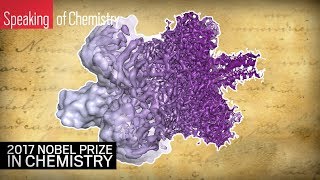
The 2017 Nobel Prize in Chemistry: Cryo-electron microscopy explained
Jacques Dubochet, Joachim Frank, and Richard Henderson have claimed the 2017 Nobel Prize in Chemistry for their development of cryo-electron microscopy.
CEN Online
Virtual Tour: Electron Microscopy Imaging
A virtual journey through the Allen Institute for Brain Science's Electron Microscopy (EM) imaging program.
Allen Institute
#microminute 23 pollen under scanning electron microscope
Once again I get a chance to remotely operate a scanning electron microscope, this time looking at pollen grains. For light microscopy of the dandelion check ...
Marty Jopson
Looking at Molecules: The electron cryo-microscopy revolution at The MRC LMB
LMB scientists have been and continue to be at the forefront of the field of electron cryo-microscopy, developing a technique which allows biological molecules ...
MRC Laboratory of Molecular Biology
Introduction to the Scanning Electron Microscope (SEM)
Nanotechnology: A Maker's Course Introduction to the Scanning Electron Microscope (SEM) Link to the full Coursera course: ...
Duke University - SMIF
2.3.3 Identify structures from electron micrographs of liver cells
You are expected to be able to identify free ribosomes, rough endoplasmic reticulum (rER) lysosomes, golgi apparatus, mitochondrion and nucleus on an ...
Stephanie Castle
ZOOM 400,000x of our Bones reveal mineral crystals (Electron Microscope)
Core Strength: Extreme "Close-Ups" May Help Explain Why Our Bones Are So Strong Snapshots taken at roughly 400000x zoom reveal mineral crystals and ...
Literatures Center
50 Images Taken with a Scanning Electron Microscope
50 Images Taken with a Scanning Electron Microscope. Subscribe to The Cryptic Compendium now for more videos on a variety of subjects. Focussing mainly ...
Cryptic Compendium
Bringing Science Closer to Children through the Electron Microscope
Hitachi High-Tech is a world-renowned manufacturer of electron microscopes. In aims to boost children's interest towards science, the company has been ...
CATCH JAPAN
WHY YOU CAN’T SEE THE CORONAVIRUS UNDER A MICROSCOPE | Electron Microscopy Explained by a Teenager
In this video, I explain why you cannot look at the coronavirus under a basic light microscope. I explain the basics behind electron microscopy and how it works.
Microbial Luke
Thwarting the next viral onslaught using electron microscopy | Dmitry Lyumkis | TEDxSanDiegoSalon
Our visible world is composed of microscopic cells, but inside each cell there's another world, orders of magnitude smaller still, a world of molecules. Within this ...
TEDx Talks
1.2 Skill: Interpretation of electron micrographs
Interpretation of electron micrographs to identify organelles and deduce the functions of specialized cells. Electron micrographs used with full permissions and ...
Stephanie Castle
Ultrastructure of Smooth Muscle Electron Microscopy in Biology and Medicine
Samuel McCombs
Brain Circuits: Harvard Medical School Researchers Crawl a Neural Network
Scientists can finally look at circuits in the brain in all of their complexity. How the mind works is one of the greatest mysteries in nature, and this research ...
Harvard Medical School
Zooming in on the Human Brain
A visually stunning tour of the human brain -- from anatomy to cells to genes and back.
Allen Institute
What's the smallest thing the human eye can see?
Our eyes are limited, but our microscopes are incredibly powerful. Watch this 90-second tour of the tiny things at the edge of human vision. Subscribe to our ...
Vox
Electron microscope images ||Top 15 Best Electron microscope images || stunning microscope images
Top 15 stunning Electron microscope images Seeing the virus up close helps us understand it. best Electron microscope images which will stun you..human cell ...
Fun with Fundamentals
Scanning Electron Microscope facility at the University of Derby
Impressive magnification is powering advances in areas such as technology, industry and research - our scanning electron microscope (SEM) can magnify ...
University of Derby
Eva Nogales (UC Berkeley): Introduction to Electron Microscopy
https://www.ibiology.org/techniques/transmission-electron-microscopy/ Transmission electron microscopy (TEM) offers the possibility of visualizing biological ...
iBiology
Electron Microscope
Electron Microscope (i). Basics of Electron Microscope (ii). Advantages over ordinary optical microscope (iii). Applications.
Physics Life
2014 Microscopy & Microanalysis - Radiolysis during Liquid Cell Electron Microscopy
Radiolysis during Liquid Cell Electron Microscopy Nicholas M. Schneider1, Michael M. Norton1, Brian J. Mendel1, Joseph M. Grogan1, Frances M. Ross2, and ...
Nicholas Schneider
Cryo Electron Microscopy: Revolutionizing the world of structural biology and healthcare
Presented At: Gibco ExpressionWorld 2018 Presented By: Marc Storms - Product Marketing Manager Life Sciences Business Unit, Materials and Structural ...
LabRoots
Hitachi HT7800 Series 120 kV Transmission Electron Microscopes (TEM)
The HT7800 series feature modern 120 kV transmission electron microscopes (TEM) with multiple lens configurations, including a standard lens for ...
HitachiHTA
What is Cryo-Electron Microscopy (Cryo-EM)?
http://www.ucsf.edu/news/2015/05/129836/resolution-revolution-building-better-microscope-see-atomic-level There are thousands of different kinds of proteins ...
UC San Francisco (UCSF)
Electron Microscopy of the Lung Lung Biology in Health and Disease
Patricia Stone
PSW 2420 Single Particle Cryo Electron Microscopy | Joachim Frank
Lecture Starts at 13:50 PSW #2420 February 21, 2020 Single Particle Cryo-Electron Microscopy Joachim Frank, 2017 Noble Prize in Chemistry ...
PSW Science
50 Amazing Things Under Electron Microscope [ SEM Images ]
50 Amazing Things Under Electron Microscope SEM Images In this video you can see 50 amazing that are seen and captured under electron microscope.
Top Bucket
Osaka University - Research Center for Ultra-High Voltage Electron Microscopy
The development of ultra-high voltage electron microscope and materials and bio-science application of electron microscopy at Osaka University The research ...
WebsEdge Science
Human Body Parts Under Electron Microscope!!
In this video I'm going to show you human body parts under electron microscope. Why not subscribe it will take only a second!! Thanks for watching!! Have a nice ...
Nami Experiments
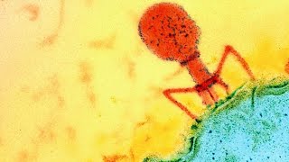
Virus electron microscope images - RNA viruses - virology
Web site : - https://medilabzone.com Hey friends I'm medical laboratory scientist.This video has information about Virus electron microscope images - RNA ...
MEDI LAB ZONE
Lecture "Cryo-Electron Microscopy Method in Structure Biology" by Ping Zhu in SibFU
Сибирский федеральный университет
What Does Cancer Look Like? | Cancer Research UK
What does cancer look like? Find out how different cancer cells appear with this video from Cancer Research UK. The video is a montage of images taken by the ...
Cancer Research UK
Renovo Neural 3-dimensional electron microscopy of Neuronal cell body
renovoneural
Introduction to In Situ Correlative Light and Electron Microscopy CLEM
Dr. Sangeetha Hari from DELMIC explains what correlative light and electron microscopy (CLEM) is and the benefits of the unique in situ SECOM system for cell ...
AXT
Team Talk: Electron Microscopy | Showcase 2017
Researchers from the Allen Institute Electron Microscopy Team present on "Mapping micro-connectivity and cell types using high throughput electron ...
Allen Institute
Shining Light on Nanomaterials with Optically Coupled Electron Microscopy Webinar
Light is a ubiquitous and critical resource in our daily lives. Light facilitates our ability to see, enables near-instantaneous global communications, and is the sole ...
Gatan, Inc.
Electron Microscope Images Of Pollen Grains | Electron Microscope Images | Photos | Pictures
Related Video : Electron Microscope Images Of Viruses With Names | Top 10 Viruses Diseases With Images And Names https://youtu.be/sDE2UV_boA8 Top 30 ...
Wonder Life Click
BIOLOGY 10 - Basic Microscope Setup and Use
This program is designed as a basic tutorial for students enrolled in Biology 10 who are first learning to setup and use lab microscopes. Produced by Technology ...
Fresno State
ZEISS Webinar: Enabling Connectomics with Multi-beam Scanning Electron Microscopy
In this webinar by ZEISS and Bitesizebio will outline some of the challenges in Connectomics and explain recent technological developments. Focus will be ...
ZEISS Microscopy
Motor proteins caught “swinging on monkey bars”
Dr Stan Burgess, of the University of Leeds's Faculty of Biological Sciences, explains research published in the journal Nature Communications that gives us our ...
University of Leeds
Virus electron microscope images - DNA viruses - virology
Hey friends I'm medical laboratory scientist.This video has information about Virus electron microscope images - DNA viruses - virology. I make this video with ...
MEDI LAB ZONE
How to Use an Ion Gun Under the Scanning Electron Microscope
How to use an ion gun in scanning electron microscopy studies? In this video we will demonstrate how a focused ion beam (created with an ion gun) can be ...
Captain Corrosion